How do we decide to move?
Overview
Complex movements arise from the concerted action of many neural circuits distributed across the brain that combine sensory information, memories of past experiences, and internal state. In contrast, simpler reflexive actions can be driven by short neural pathways that reside entirely in the brainstem or spinal cord. In between these two extremes of behavioral complexity are actions that may be driven by different neural pathways in different contexts. The Machado lab uses a combination of new multiregion neural recording techniques, tissue clearing methods, and computational data analysis to study how motor command pathways are recruited during these kinds of behaviors. The results from our work will lead to a better understanding of how neural computation produces behavior—and will inform new approaches for treating neurological conditions and for building brain machine interfaces.
How do motor command pathways interact to generate specific movements?
Past studies have shown that many parallel pathways that arise in different regions across the brain are each involved in driving specific movements. But until recently, it was difficult to study how these parallel pathways interact with each other on a moment-to-moment basis to produce behavior. This is because it was not straightforward to simultaneously record from large numbers of neurons in distinct regions. Thanks to recent advances in neural recording technology, we are now able to simultaneously measure the commands sent by different brain areas during different kinds of movements. This lets us study how motor command syntax might change as a function of changing brain state (e.g. with thirst or arousal), or as a consequence of neurodegenerative disease.
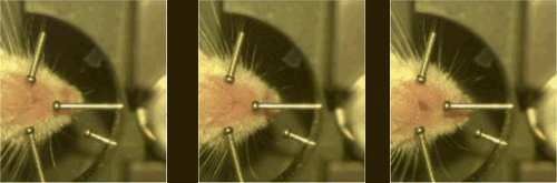
Above: A mouse licking in different directions as viewed from below. From: Kauvar* and Machado*, 2020
We study brainwide circuits using brainwide recording techniques.
Optoencephalography (widefield Ca2+ imaging and COSMOS)
We use COSMOS, a single-photon imaging technique, to measure neural activity from across the curved surface of mouse dorsal cortex.
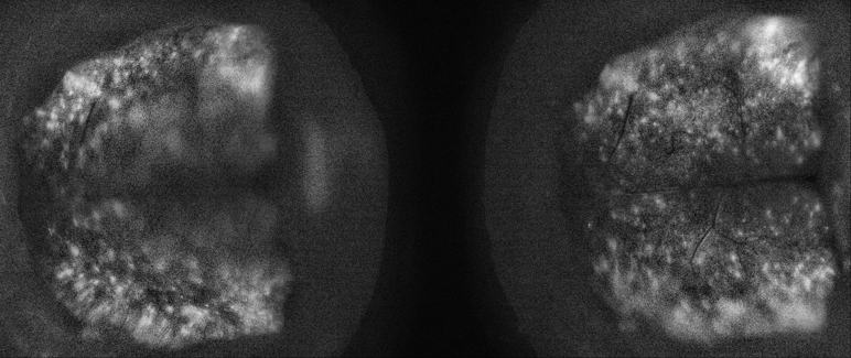
Above: Representative data obtained on a COSMOS microscope. From: Kauvar* and Machado*, 2020
Large-scale electrophysiology (Neuropixels)
We use multielectrode arrays called Neuropixels to simultaneously record from hundreds of neurons.
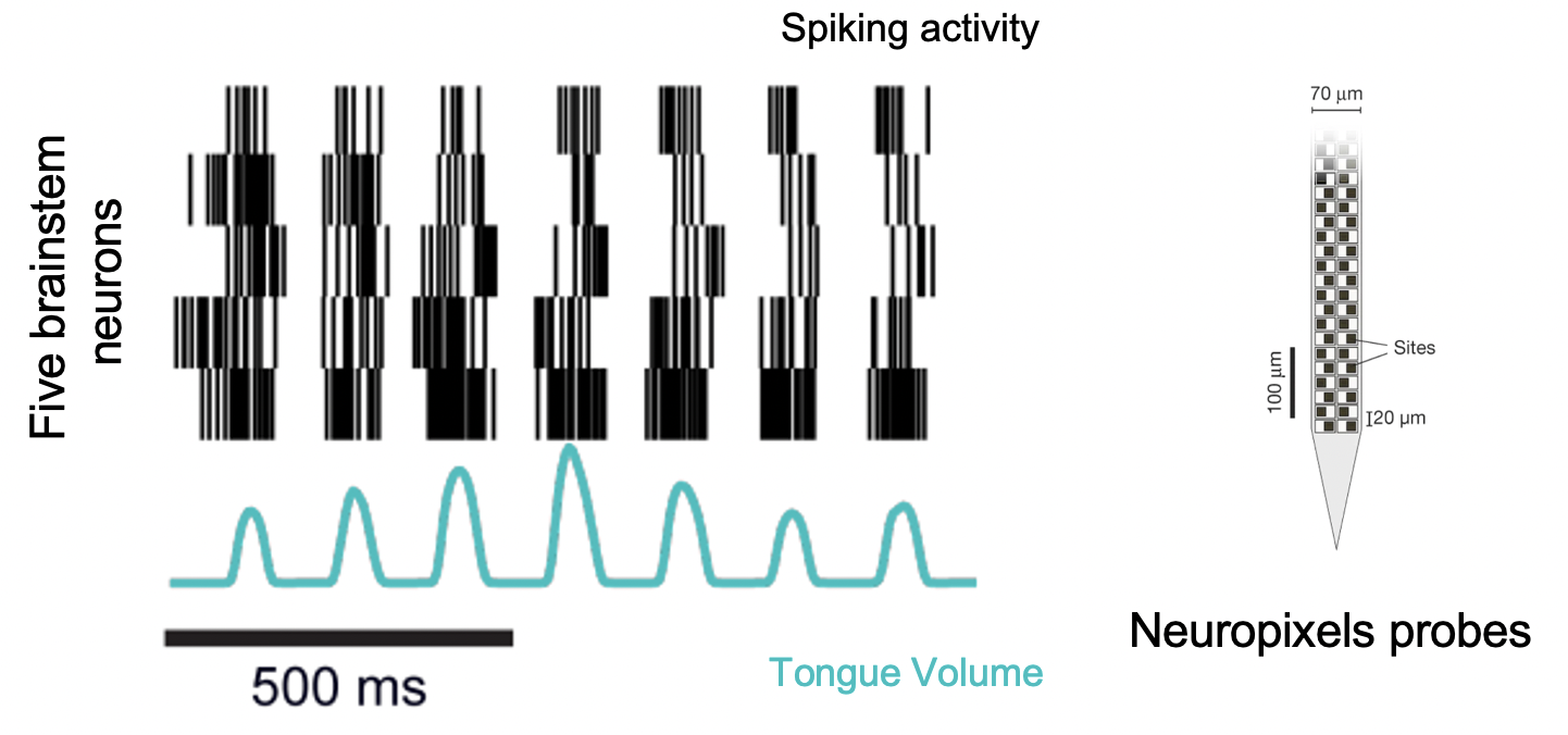
Above: Spiking activity from single units in the medulla. Panel on right adapted from Jun et al. 2017
Tissue clearing techniques and light sheet microscopy
We use tissue clearing techniques like SHIELD and DISCO to perform whole-brain histology and to reconstruct electrode tracks.
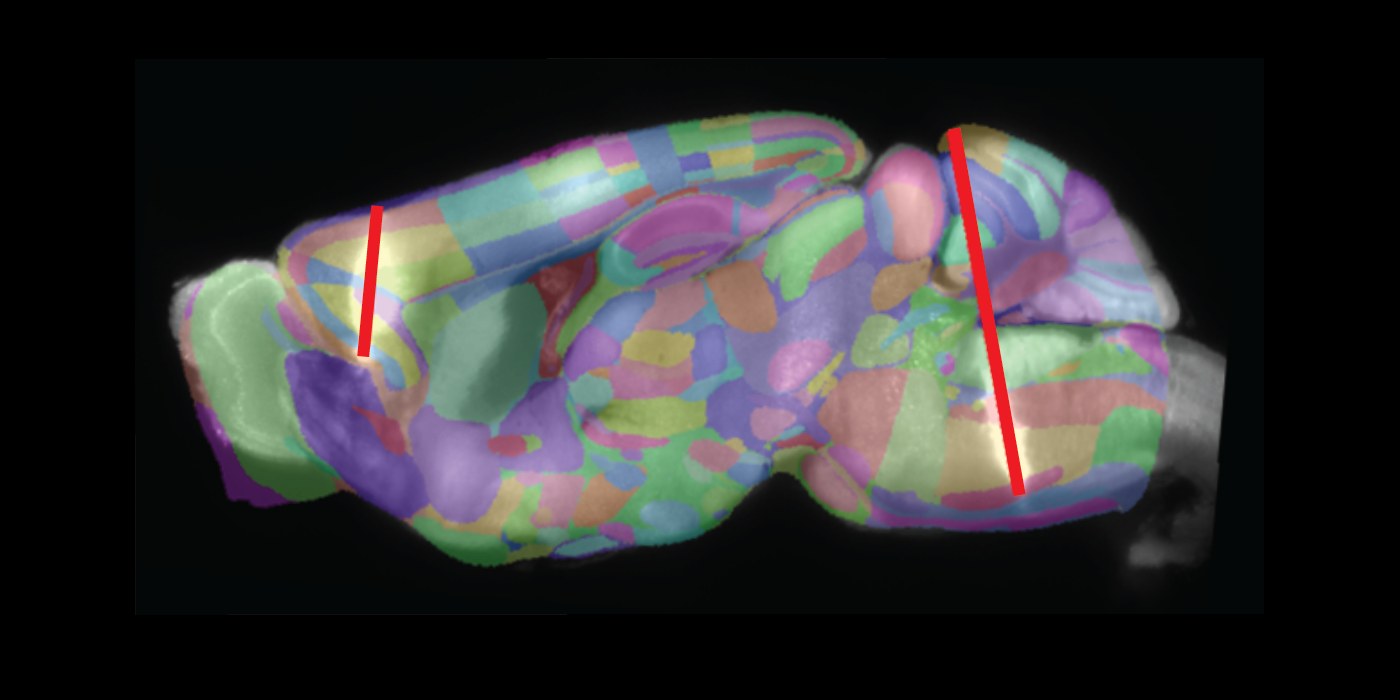
Above: Fluorescent Neuropixels electrode tracks aligned to the Allen Brain Atlas.
Analysis of neural population dynamics
We use and develop new algorithms and model-based approaches for analyzing the large-scale neural datasets that we collect.